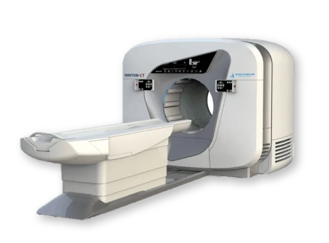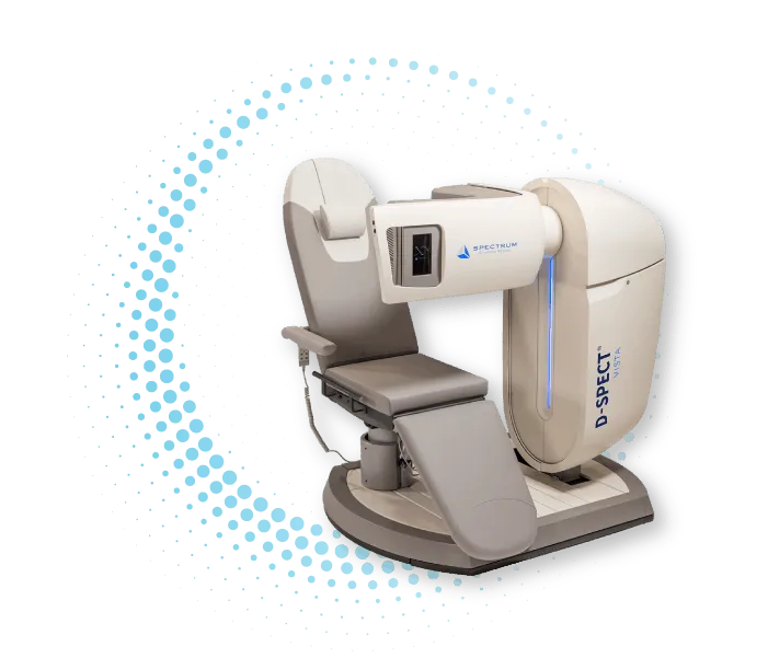
D-SPECT Series nuclear cardiology camera
A high-performance cardiac SPECT system that combines solid-state detector technology with advanced quantitative analysis algorithms. Designed for cardiovascular imaging, the system supports high-quality image acquisition, fast scanning workflows, and improved patient comfort.
Key feature: solid-state CZT detectors
The D-SPECT Series is based on solid-state detectors made of cadmium zinc telluride (CZT). Compared with conventional NaI detectors, CZT technology provides higher sensitivity, energy resolution, and photon detection efficiency.
This architecture supports fast data acquisition in cardiac SPECT studies and enables advanced quantitative analysis methods.

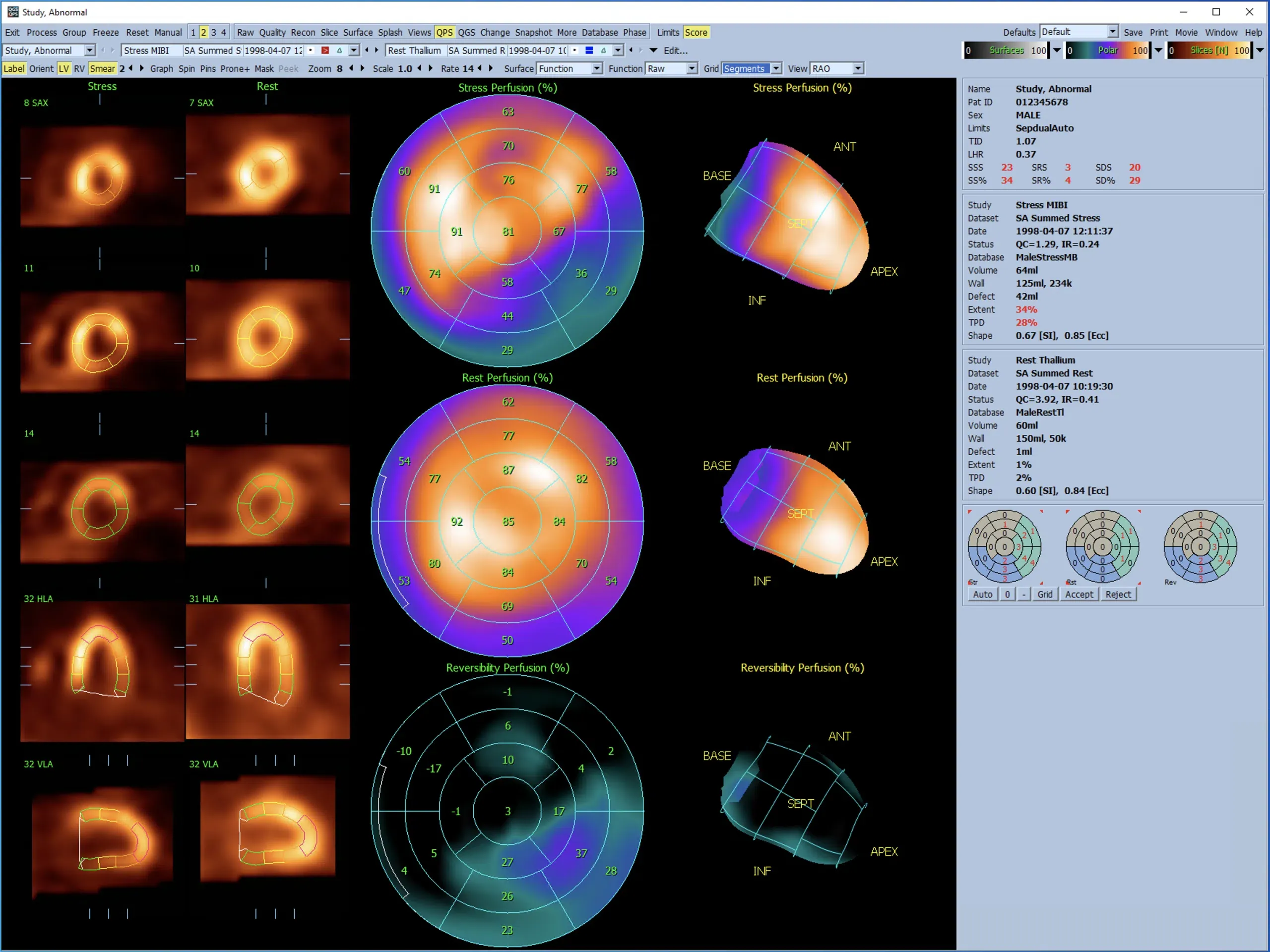
Clinical capabilities and diagnostic imaging
The system is designed for nuclear cardiology and supports myocardial perfusion imaging, quantitative analysis, and studies in patients with different anatomical characteristics.
- Quantitative assessment of myocardial blood flow: dynamic SPECT scanning can be used to calculate MBF and MFR, including coronary flow reserve assessment. This may be useful in the diagnosis of multivessel and microvascular disease.
- Focused region-of-interest scanning: the design of the detector columns enables targeted scanning of the myocardial region and improves useful signal detection efficiency.
- Imaging of patients with high BMI: the hardware configuration and tungsten collimators help acquire diagnostic images in patients with increased body weight.
- Multi-isotope imaging: the energy resolution of CZT detectors supports signal separation when working with multiple isotopes, if this mode is supported by the protocol and software.
Patient safety and comfort
The system design is intended to reduce examination time, decrease the need for repeat scans, and improve patient comfort during the procedure.
- Reduced radiation exposure: the high sensitivity of CZT detectors may allow protocols with reduced radiopharmaceutical activity while maintaining diagnostic image quality.
- Fast scanning: shorter data acquisition time may reduce patient fatigue and decrease the likelihood of motion artifacts.
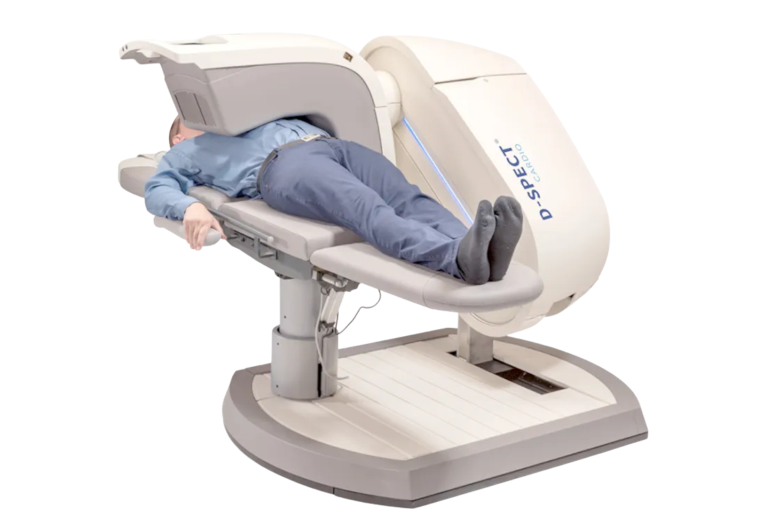
- Open architecture without gantry rotation around the patient: the detectors move inside the housing, with no external gantry rotation around the patient. This design reduces the risk of mechanical contact and may be more comfortable for patients with claustrophobia.
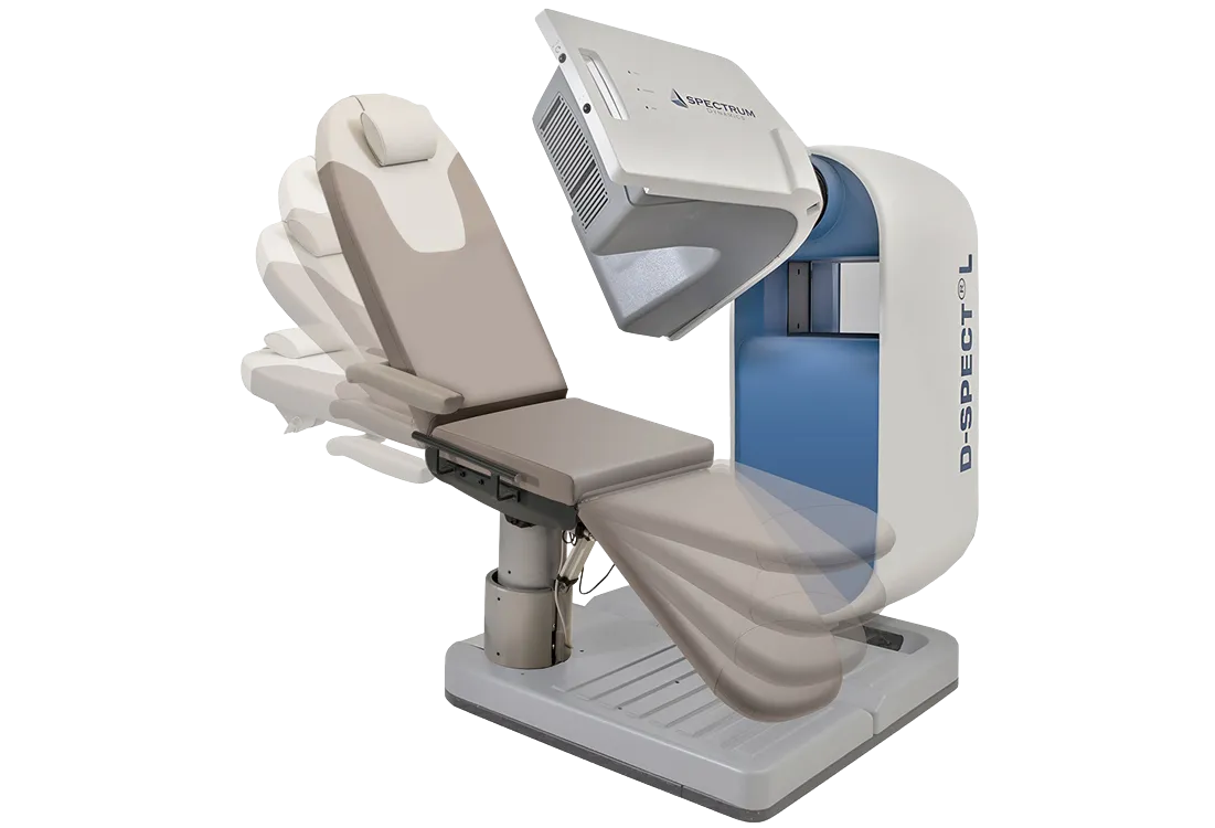
Ergonomics and design features
The gantry and integrated chair design allow patient positioning to be adapted to the clinical task and examination requirements.
- Convertible chair-table: the patient can be positioned upright, semi-recumbent, horizontal, or in an intermediate position. Scanning in different positions may be used to reduce the effect of attenuation artifacts.
- Increased load capacity: the reinforced chair structure is designed for imaging patients with increased body weight, within the parameters specified by the manufacturer.
- Free arm positioning: eliminating the need to place the left arm behind the head may improve patient comfort and reduce the risk of involuntary movements.
- Compact installation: the small footprint facilitates system placement in rooms with limited space.
Software and data processing
The D-SPECT Series hardware platform is complemented by software tools for reconstruction, post-processing, and quantitative data analysis.
- Iterative reconstruction: reconstruction algorithms based on OSEM are used to generate images and improve visualization quality.
- Integration with quantitative analysis software: the system may support operation with INVIA 4DM, Cedars-Sinai QGS/QPS, and Syntermed Emory Toolbox, if the corresponding integration is included in the supplied configuration.
- List Mode data acquisition: List Mode allows events to be stored for subsequent regrouping of static and dynamic series data without rescanning the patient.
Workflows and benefits for the clinic
The D-SPECT Series is designed to optimize routine workflows in the nuclear medicine department and improve staff efficiency.
- Control near the patient: the touchscreen integrated into the detector area allows the operator to perform positioning and settings next to the patient.
- Integration with medical information systems: DICOM support enables data exchange with PACS and other in-hospital systems.
- Remote service support: the system may support remote diagnostics and service maintenance, if provided by the service configuration.
- Quality control: daily setup and quality control procedures are performed in accordance with the manufacturer’s protocol.
Target patient groups
The system can be used for cardiac SPECT studies in various patient groups, including:
- patients with diabetes mellitus;
- patients with chronic kidney disease;
- patients with cardiomyopathy and heart failure;
- elderly patients;
- patients with chest pain and normal coronary angiography results;
- patients with severe obesity.